We have discovered a protein, termed the CAncer REcognition (CARE) antigen (Ag), which binds a CARE Antibody (Ab) from cancer patients' sera but very little in healthy volunteers, patients with a history of cancer or benign conditions. The antibody is of the IgM primitive form and does not seroconvert to IgG. Data show that cancer patients have elevated amounts of this unique IgM antibody Ab directed against the CARE antigen Ag. This may be the first time a cancer antigen can be quantitated along with the immune complex and free Ab.
The OD readings (absorbance) readings for a variety of negative, normal plasma used from the lyophilized commercial source and positive controls were measured. The serums could be stored at 4 C for up to 206 days with little change in the absorbance readings.
Healthy Volunteers - CARE Ab Test. Mean Relative Concentration (MRC) - (Mean ± SD).
Cancer Patient MRC Values.
False Positive - False Negative Percentages CARE Ab Test.
Patient AS - Colon Cancer with a staging of C2 secondary to Stage 3.
CEA negative.
Patient LB - Primary Breast Cancer T2N0M0,
Diploid S-phase = 4%, ER/PR+
The beauty of having a short-lived IgM as the marker for cancer is that it stands to reason that when the tumor is removed completely, the CARE antibody should also decrease. Therefore, the CARE antibody can be used to monitor residual disease.
| Enzyme-Linked Immunosorbent Assay (ELISA) | |
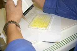
The patient's serum is diluted 1:100 (YES, the test is that sensitive), and 100 ul is added to each well in duplicate for the sample determination. Note the pentameric IgM binding specifically to the CARE Antigen (CA), not to other proteins (red) or BSA. The next step is to simply detect human IgM binding to the plate well bottom. This is accomplished using a commercial antibody produced in a goat against human IgM which has an enzyme called horse radish peroxidase (HRP) attached to it. After one hour incubation at room temperature, the plate is washed a final time, and a colorless dye is added. The dye is oxidized by the HRP to a blue color during a 10 min. incubation at room temperature. Subsequently, 1 N sulfuric is added to each well with a 12 place multipipetor to stop color development. This converts the blue to a yellow color.
The intensity of the yellow color (absorbance) is directly proportional to the amount of HRP present, which is directly proportional to the IgM in each well. In other words, the determination of the quantity of IgM in each well is directly proportional to the absorbance of the yellow color at 405 nm in a microtiter plate reader.
A positive control serum is used to develop a standard curve from which the absorbance of the patient unknown is read. The positive control serum (after dilution 1:100 as with all serums) is serially diluted with commercially available normal human plasma (lyophilized and made fresh) to generate an eight point curve which is analyzed using the standard four parameter curve analysis. The Mean Relative Concentration (MRC), i.e., relative to the standard negative plasma, is determined from the curve by comparisons of the absorbance readings of the unknown patient sample (in duplicate) with the standard curve (data points are means of duplicate measurements). The MRC values are presented as whole integers by multiplying the results by 1000.
EACH plate contains its own standard curve allowing 36 CARE Ab determinations from 36 patients to be determined in duplicate per plate.
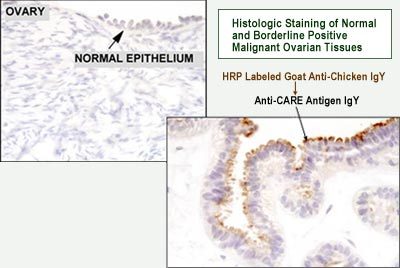
| The Antigen Assay is similar except an Ab raised in chickens is attached to the plates for "trapping out" Ag from the serum. Purified, pooled human IgM from cancer patients is added to attach to the Ag with subsequent IgM detection as with the Ab assay above. | |
Following are videos clips showing carcinoma cells that were cultured and prepared for confocal microscopy:
A three-dimensional rotational film of three prostatic cells (LNCaP) stained with anti-LT11 and a Hoescht 33258 nuclear counterstain. Krypton lasers were used for imaging purposes due to the AlexaFluor 594 secondary antibody. The film reveals very specific staining of the cell membrane as well as some filamentous staining. Again, emphasizing that LT-11 may be a portion of an intermediate filament.
The same prostatic cancer cells but showing a step by step movement through the cells using a 0.6 µm step-size.
A three-dimensional rotational film of ovarian carcinoma cells (OVCAR3) stained with anti-LT11, again emphasizing surface staining and some filamentous cortical staining.
The same ovarian carcinoma cells with the additional Hoescht 33258 nuclear counterstain.
The same ovarian carcinoma cells but showing a step by step movement through the cells using a 0.5 µm step-size.
Monday
9:00 am - 6:30 pm
Tuesday
9:00 am - 6:30 pm
Wednesday
9:00 am - 6:30 pm
Thursday
9:00 am - 6:30 pm
Friday
9:00 am - 6:30 pm